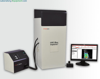
- UVP iBox Explorer 2 live cell in vivo Imaging Microscope with Optichemi 695 camera for macro to micro fluorescent small animal imaging
- Images organs and cells subcutaneously and within body cavity of living mice
- Parcentered and parfocal optical configurations for seamless transition from macroscopic to microscopic scale
- Visualizes micro injection of cancer cells and easily detects GFP/RFP and other fluorescent markers in small animals
- Magnification range: seamless navigation from 0.17x to 16.5x
Gel Doc Systems
UVP iBox Explorer 2 Imaging Microscope by Analytik Jena
Read more
Manufactured By: Analytik Jena
Warranty: Three-Year Manufacturer Warranty; One-Year Warranty for Camera
Print
Want a quote quickly ?
Submit a QuickQuote requestProducts similar to this one
-
 Group of 4 productsUVP iBox Studio Touch In Vivo Fluorescence Imaging System by Analytik Jena FromAs low as $59,000Some ship in 20 - 25 days
 - Usually Ships in 20 - 25 days
- Group of 8 productsUVP ChemStudio Western Blot Imaging Systems by Analytik Jena FromAs low as $38,140Some ship in 6 - 10 days
- Group of 8 productsUVP GelStudio DNA Gel Documentation Imaging Systems by Analytik Jena FromAs low as $16,282Some ship in 6 - 10 days
- Group of 14 productsUVP GelSolo UV Gel Documentation Systems by Analytik Jena FromAs low as $12,718Some ship in 6 - 10 days
Page 1 of 2
Other products you may find relevant
- Some ship in 6 - 8 days
- Some ship in 3 - 5 days
- Some ship in 5 - 7 days
- Some ship in 2 - 4 days
Page 1 of 1
Features and Benefits

Critical Environment Solutions
Terra's mission is to help customers in highly regulated industries transform the world with critical environment solutions that improve health, safety, performance, and yields. These environments may comply with stringent UL, ISO, IEST, ASTM and OSHA standards and local requirements.
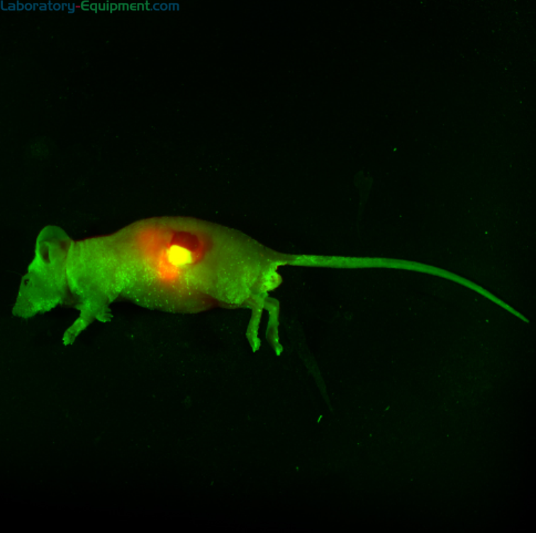
Fluorescent Cancer Cell Detection
Zoomed view of HT1080 fluorescent cancer cells injected into the epigastrica cranialis immediately shows a cluster of cancer cells escaped around injection site and in the bloodstream
Products similar to this one
-
 Group of 4 productsUVP iBox Studio Touch In Vivo Fluorescence Imaging System by Analytik Jena FromAs low as $59,000Some ship in 20 - 25 days
 -
 Usually Ships in 20 - 25 days
 -
 Group of 8 productsUVP ChemStudio Western Blot Imaging Systems by Analytik Jena FromAs low as $38,140Some ship in 6 - 10 days
 -
 Group of 8 productsUVP GelStudio DNA Gel Documentation Imaging Systems by Analytik Jena FromAs low as $16,282Some ship in 6 - 10 days
 -
 Group of 14 productsUVP GelSolo UV Gel Documentation Systems by Analytik Jena FromAs low as $12,718Some ship in 6 - 10 days
Page 1 of 2
Other products you may find relevant
-
 Some ship in 6 - 8 days
 -
 Some ship in 3 - 5 days
 -
 Some ship in 5 - 7 days
 -
 Some ship in 2 - 4 days
Page 1 of 1