Learn More: SpotWare Scanners
Specifications for SpotWare Colorimetric Scanner
| Instrument Size | 10.63W x 18.11D x 4.53H in. (270 x 460 x 115 mm) |
|---|---|
| Adjustable Scanning Resolutions | 5, 10, 20, 50 µm |
| Operating System | Microsoft Windows XP |
| Scan Area | 25 x 76 mm with separate barcode detection capability |
| Scan Speed | 1.0 cm2 in 30 seconds at 10µm resolution |
| Total Dynamic Range | >7 logs (10,000,000 fold) |
| Standard Tiff Intensity Value Range | 0-65.536 |
| Image Format | 16-bit TIFF file format with 065,536 intensity values |
| Electrical | 110V, 50-60Hz |
| Applications | Research and diagnostics, DNA, RNA, protein, antigen, and antibody microarrays, chromatic detection of microarrays, western blots, microwestern arrays |
| Operating System | Windows XP |
| Additional features |
|
| Product Contents |
|
Adjustable Resolution Settings of SpotWare Colorimetric Scanner
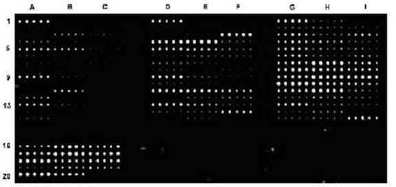
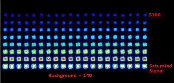
 Microarray instruments work in conjunction to manufacture, detect and scan samples for quantitative detection analysis. The images below demonstrate how the SpotWare™ Colorimetric Microarray Scanner functions with other microarray instruments as well as showcasing the versatility of the SpotWare’s four resolution settings.
The above image represents the process of creating, detecting and scanning a microarray when conducting quantitative detection analysis. Protein microarray was manufactured using the ArrayIt® Protein Microarray Platform in conjunction with ArrayIt® SpotWare™ Colorimetric Microarray Scanner for detecting. By diluting antibodies in 1X Protein Printing Buffer, the antibodies were printed in quintuplicate onto SuperProtein Substrates using a microarray spotter incubated with patient sera, stained with a secondary antibody conjugated to Alkaline Phosphatase (AP), and developed with an AP Developing Kit prior to scanning at a gain setting of 1.0 and 10 µm Resolution.
 Sample 16-bit image was acquired with the SpotWare™ Colorimetric Scanner. Presented in a rainbow palette at a resolution of 5µm clearly demonstrates the 1 x 10⁷ dynamic range of the SpotWare.
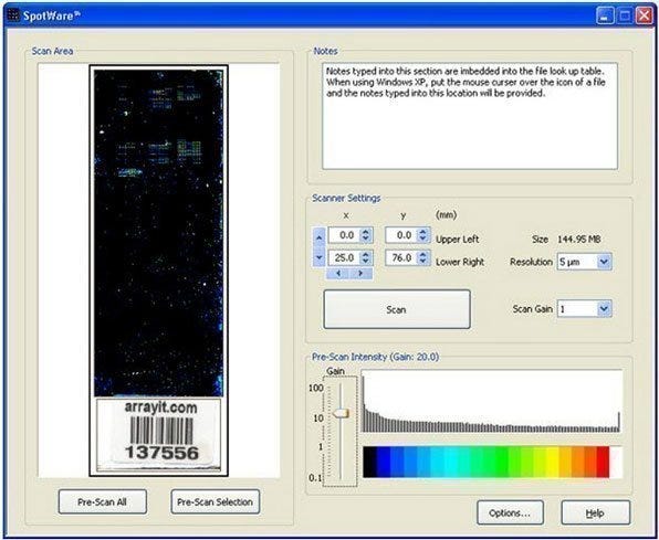
Main Software Window and Graphical User Interface
 The scanned substrate image (left) with data obtained using alkaline phosphatase (AP) labeling is coded to a rainbow palette for easy viewing. Users have the choice to pre-scan all or a selection of the image. There is a text window (top right) for inputting experimental notes. The scanner settings (middle right) with a scan button is where users can input: scan area coordinates (x, y), file size (MB), scan resolution (µm) and scan gain (0.1100). The pre-scan intensity window (bottom right) indicates the scan gains and an intensity histogram coded to a rainbow palette.
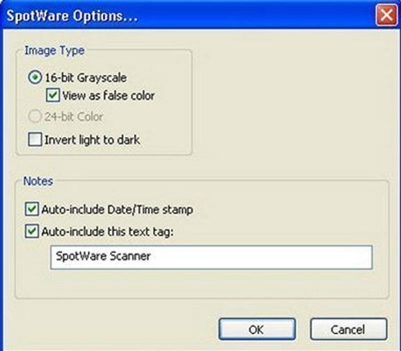
SpotWare Options Window
 Using the Arrayit Alkaline Phosphatase (AP) Kit (#CAT APK) to develop this microarray, this Pre-Scan Selection image is at a scan gain of 7.0 with a visible increase on signal intensity compared to Figure 7. while the background noise remains low.