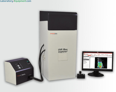
- UVP iBox Explorer 2 live cell in vivo Imaging Microscope with Optichemi 695 camera for macro to micro fluorescent small animal imaging
- Images organs and cells subcutaneously and within body cavity of living mice
- Parcentered and parfocal optical configurations for seamless transition from macroscopic to microscopic scale
- Visualizes micro injection of cancer cells and easily detects GFP/RFP and other fluorescent markers in small animals
- Magnification range: seamless navigation from 0.17x to 16.5x

UVP iBox Explorer2 with Optichemi 695 camera detects fluorescence markers in vivo and easily transitions from macroscopic to microscope scale (PC not included) | 1017-PP-07 displayed
Gel Doc Systems
UVP iBox Explorer 2 Imaging Microscope by Analytik Jena
Read more
Manufactured By: Analytik Jena
Warranty: Three-Year Manufacturer Warranty; One-Year Warranty for Camera
Summary
- UVP iBox Explorer 2 live cell in vivo Imaging Microscope with Optichemi 695 camera for macro to micro fluorescent small animal imaging
- Images organs and cells subcutaneously and within body cavity of living mice
- Parcentered and parfocal optical configurations for seamless transition from macroscopic to microscopic scale
- Visualizes micro injection of cancer cells and easily detects GFP/RFP and other fluorescent markers in small animals
- • Magnification range: seamless navigation from 0.17x to 16.5x
- iBox Explorer 2 bioimaging system used in: multiplex in vivo, cancer research, gene expression, nanomedicine, near IR, immunology, heart disease, alzheimer’s, plant and agriculture
- Features:
- Superior cooled CCD camera and optics optimized for VIS – NIR imaging
- Motorized optics enable viewing whole animal to single cell level
- Upright optics provide ultra long working distance
- Detailed fluorescent in vivo imaging and high numerical aperture (NA)
- Software interface with eLITE excitation filters and light intensity
- Viewing window for quick sample inspection
- Uniform temperature maintained by slide-out warming plate
- Configurable filters for fluorescence microscopy enable generation of continuous excitation spectrum
- Provides optimum imaging conditions with light tight darkroom
- Adjustable position of motorized X, Y, Z stage with joystick
- Enables in vivo research study from whole organs to single cell
- Quickly detects fluorescence
- Specifications:
- Darkroom Dimensions: 17.5"W x 19.5"D x 41"H (44.5 x 49.5 x 104 cm)
- eLITE Dimensions: 12.5 x 13.5 x 10 in. (31.8 x 34.3 x 25.4 cm)
- Emission Filters with four-position interchangeable rack: 515 nm Longpass, GFP, RFP, Neutral Density; Additional wavelength filters available
- Controls: Automated, software controlled
- Travel: Precision motorized X, Y, Z
- Resolution: X = 10 μm, Y = 10 μm, Z = 1 μm
- Position/Focus: Controlled by joystick/software
- Warming Plate: 37°C, user adjustable
- Light Sources: 150 watt Xenon
- Fiber Optic: Coaxial and epi illumination with light heads mounted to the darkroom
- Excitation Filters: Eight position wheel includes filters - GFP, RFP (others available)
- Controls: Via software
- Camera OptiChemi 695:
- Monochrome
- 16 bit
- 2184 x 1472 pixel Resolution
- 3.2, extendable to 9.6 Megapixels
- Cooling: -60°C from ambient
- Peak QE: 88% (quantum efficiency)
- Software(requires Microsoft Windows 7,8, or 10 (32-bit or 64-bit):
- Interface: Camera, objectives, darkroom, eLITE
- Tools: Macros and templates
- Analysis: Extensive functions including area density measurements, volume measurements and tumor sizing
- Documentation: Create reports and export data
- Compliance: Supports 21 CFR Part 11
- Optical magnification: 0.17x, 0.25x, 0.5x, 1.66x, 2.5x, 4.5x, 7.5x, 8.8x, 16.5x
- FOV Observation Range (mm): 90 x 90, 60 x 60, 30 x 30, 9 x 9, 6 x 6, 3.3 x 3.3, 2 x 2, 1.7 x 1.7, 0.9 x 0.9
- Optional UVP anesthesia kit for immobilizing small animals #98-0098-01

