
What is Gel Electrophoresis?
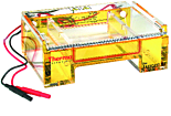
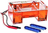
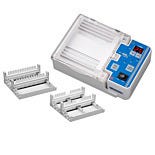
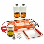
Gel electrophoresis systems utilize a porous, electrically-charged gel matrix to separate distinct nucleic acid and protein sequences based on molecular weight (or fragment size). The gel box is designed to include a cathode, or negatively-charged electrode, at one end of the medium and an anode, or positive-charged electrode, at the opposite end.
Electrophoresis Protocol
The gel box is filled with an ionic buffer that creates an electrical field when the cathode and anode are connected to power. As proteins, DNA and RNA molecules carry an intrinsic negative charge; the fragments migrate through the gel medium toward the anode. The speed of migration correlates to the size of the fragment; smaller molecules will move faster through the porous gel than larger, slower molecules. Once completed, the gel run results in unique bands of nucleic acids or proteins separated by molecular weight. Compared against a positive control ladder for reference, the appropriate fragments are excised from the gel for further purification.
A - Electrophoresis Gel Configuration
(back to chart)
A1 - Electrophoresis Systems
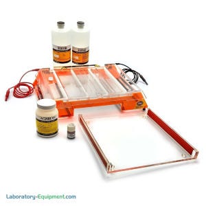
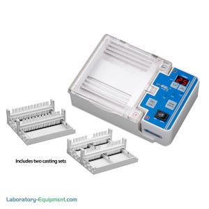
Horizontal Electrophoresis Gels cast in a horizontal gel orientation are used primarily for nucleic acid separation within an agarose matrix. As the pores within an agarose gel are larger than the pores within a polyacrylamide gel, the DNA and RNA molecules, which are larger than protein molecules, are better suited to migrate effectively through agarose. Since horizontal systems expose the gel matrix to atmospheric oxygen, agarose is chosen as the preferred medium over polyacrylamide, which does not polymerize in the presence of O2 gas.
View Online: Thermo Fisher Gel Horizontal Electrophoresis
A2 - Vertical Electrophoresis Systems
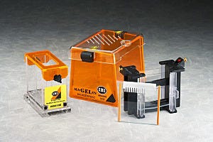
Vertical Electrophoresis Gels cast in a vertical orientation are used primarily for protein separation within an acrylamide matrix. As acrylamide pores are smaller in diameter than agarose pores, acrylamide gels yield a higher resolution and greater separation of proteins, which are smaller than nucleic acid fragments. Since vertical systems require a thinner (less than 2 mm) gel, acrylamide is the optimal choice over agarose, which contains larger gel pores that inhibit fragment migration through a thinner matrix. Learn more about the differences between horizontal vs vertical gel electrophoresis systems.
View Online: Horizontal DNA and Protein Electrophoresis Systems - IBI Scientific
B - Electrophoresis Gel Matrix
(back to chart)
B1 - Agarose Gel for DNA Electrophoresis
Agarose gels contain larger (100 to 500 nm) and less uniform pores than acrylamide gels, making agarose optimal for nucleic acid separations, which involve larger fragments than protein separations. Agarose gels achieve better fragment separation during horizontal runs, which are designed to accommodate thicker (greater than 2 mm) gels than vertical runs.
Compare Online: Agarose Electrophoresis Gels
B2 - Acrylamide Electrophoresis Gel
Acrylamide gels contain smaller (10 to 200 nm) and more uniform pores than agarose gels, making acrylamide optimal for protein separations, which involve smaller molecules than DNA and RNA separations. Acrylamide gels yield clearer fragment separation during vertical runs, which are designed to require thinner (less than 2 mm) gels than horizontal runs. Acrylamide does not polymerize, or harden, in the presence of atmospheric oxygen, making acrylamide incompatible with horizontal gel boxes, which expose the gel matrix to O2 gas.
Compare Online: Polyacrylamide Gels
C - Electrophoresis Sample Type and Resolution
(back to chart)
C1 - Nucleic Acid Electrophoresis
For higher resolution and separation clarity, horizontal-oriented agarose gels are optimal for DNA and RNA molecules. Because horizontal gels utilize a continuous buffer system, the nucleic acid fragments are accessible during the separation procedure.
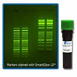
Compare: Benchmark SmartGlow Pre-stain for Nucleic Acid Gels
C2 - Serum Protein ElectrophoresisProtein
For higher resolution, vertically-oriented acrylamide gels are best suited for protein molecule separation. As vertical gels utilize a discontinuous buffer system, the buffer can only flow through the gel when moving from the top to the bottom chamber, allowing for more precise control of the voltage gradient. The optimization of the voltage gradient yields higher separation clarity, ideal for linear protein strands, which may demonstrate similar molecular weights.
D - Voltage
(back to chart)
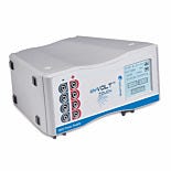
D1 - 120 Volt Electrophoresis Systems
120-volt connections are suitable for low-voltage standard residential power outlets in the US.
D2 - 240 Volt Electrophoresis Systems
240-volt connections require less current (amperage) and smaller conductors than appliances designed to operate at 120V.
E - Electrophoresis Buffer Capacity
(back to chart)
The volume and concentration of the buffer used to prepare and run the gel depends upon the application and sample type. Two commonly used buffer formulations are Tris-Acetate-EDTA (TAE) and Tris-Borate-EDTA (TBE). TAE buffer yields faster migration of linear DNA and better resolution of super-coiled or genomic DNA. TBE buffer is optimal for separation of longer DNA fragments (larger than 2 kb).
F - Electrophoresis Gel Dimensions
(back to chart)
The optimal gel box size depends upon the sample type, buffering capacity and voltage level. Smaller gel boxes are ideal for separation of short, linear DNA fragments, while larger gel boxes are optimal for longer DNA fragments (larger than 2 kb).
Where Can I Find a Trusted Supplier of Electrophoresis Equipment?
Laboratory-Equipment.com offers carefully selected electrophoresis product lines from Thermo Fisher, IBI Scientific Gel Systems, Benchmark (SmartGlow) and Accuris (myGel & myVolt). Through our worldwide network of reps, we supply some of the largest research and production facilities in the world. Laboratory-Equipment.com is a laboratory speciality division of Terra Universal. For nearly 40 years, Terra has served semiconductor, aerospace, life science, pharmaceutical, biotechnology, and medical device markets.
Shop Electrophoresis Gel and Equipment by Brand
Try before you buy!
DEMO MODELS AVAILABLE
Contact us for Demos, Samples, Brochures and more.
EMAILMon - Fri, 5:30am - 5:30pm PST


