The preparation of samples for protein and nucleic acid analysis requires two essential steps: disruption of the tissue to release individual cells and lysis of those cells to release their cellular contents. Mechanical homogenization and sonication (sometimes referred to as ultrasonic homogenization) are two mainstay techniques for these processes.
Selecting the proper technique for your particular application deserves careful thought. Each one has its benefits and drawbacks, and laboratories often choose to use these techniques in concert with one another. Keep reading for information to help select a method that avoids damaging precious or irreplaceable tissue samples.
Mechanical Homogenization
Mechanical homogenization utilizes direct physical force to bring a biological sample in solution to a state of uniform distribution, such that all fractions’ molecular composition is consistent. Traditionally, mechanical disruption was achieved by freezing tissues and then grinding with a mortar and pestle. Presently, there are two other common methods: bead-based disruption and rotor-stator disruption. These separation techniques efficiently homogenize samples, but often require further downstream fractionation to obtain the desired concentration or purity of molecules.

Beads within microtubes create the shearing effect at high speeds. Before and after of seed, tissue and plant samples. (Benchmark Scientific)
Bead-based homogenization uses plastic or metal beads combined with high-speed shaking to create shearing forces. This technique is well-suited to whole animal, insect (e.g., Drosophila melanogaster) or plant specimens, which requires disruption of sturdy cell walls. A drawback of bead-based disruption is that it requires the proper selection of bead material and diameter. For example, some plastic beads will readily bind with DNA, depleting it from solution when they are removed. Similarly, the diameter of the bead can impact the amount of force created and the degree to which a sample is disrupted. Using a bead size that is either too large or too small can result in incomplete homogenization. Thankfully, many instrument manufacturers have developed a variety of beads to suit most homogenization applications.
Rotor-stator disruption involves the use of a stationary stator housing a rapidly moving rotor. The movement of tissues and cells within the space between the rotor and stator creates a high degree of shearing force. These devices are typically hand-held and the rotor-stator attachment is interchangeable. These devices efficiently disrupt tough animal and plant tissues (e.g., muscle). One salient benefit of rotor-stator disruption is the ability to progressively increase the degree of homogenization by sequentially processing with increasingly smaller rotor-stator distance attachments. This process can effectively homogenize even the most resilient specimens. Additionally, very large-volume samples can be processed using larger disruption instrumentation. Drawbacks of rotor-stator disruption include increased cost and more cumbersome equipment management. Attachments often require washing and sterilization prior to reuse. Disposable attachments are offered, but with a higher operating cost.

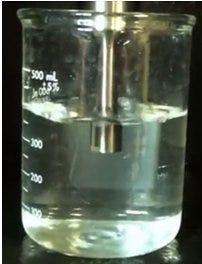
Sonicating probe in sample. (QSonica)
Ultrasonic Homogenization
Ultrasonic homogenization also utilizes mechanical forces to shear tissues and cells, however, this force takes the form of ultrasonic sound waves. This can be done either using an ultrasonic bath or a probe. This technique can be used to rapidly homogenize samples, but care must be given to the power and frequency used, as well as the duration of the processing, since improper sonication parameters can result in irreversible sample damage.
Both bath and probe sonicators utilize high-energy sound waves to disrupt samples. Probe sonication is the most frequently used process for the disruption of cells. Bath sonication is useful for large batch preparations of tissues and cells.
When starting with whole tissue, use the tissue type to determine if sonication alone is a feasible technique for effective disruption. More resilient tissues, such as muscle, require an upstream, primary disruption such as blending, snap freezing or mechanical homogenization. It is also worth mentioning that processing whole animals and some plant tissues cannot be accomplished through sonication alone. Thus, while sonication effectively and rapidly disrupts cells in solution and many tissues, it cannot be used for some starting materials.

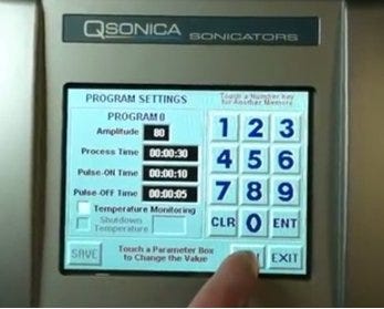
Programmable digital sonicator allows control of speed and duration. (QSonica)
Ultrasonic homogenization is a high-energy process and operators run the risk of altering the molecular make-up of the solution during sonication. Users must specify the frequency and wattage of the process. Nucleic acids, in particular, are susceptible to the shearing forces of sonication. If a sample is processed at too high of a frequency, the DNA/RNA backbone is sheared and analysis is no longer feasible. The susceptibility of nucleic acids to shearing can also benefit operators. If your desired end product is protein, users will want to remove the DNA in solution. Sonication can effectively disrupt genomic and cellular nucleic acids, so that they no longer interfere with protein purification. Lastly, the high-energy created through sonication can heat samples and result in the denaturation of proteins (if the sample is processed for an extended duration). Thus, be mindful of the duration, wattage and frequency used for ultrasonic homogenization.
The homogenization of tissues and cells is a common technique used to facilitate further downstream analysis. Myriad techniques and instruments are offered for these processes. Mechanical homogenization and ultrasonic homogenization are two commonly used techniques. Each has its advantages and drawbacks. Many times researchers chose to use the two techniques in concert in order to obtain the most desirable outcome. A summary of how they compare:
See more information about the Sonicators and Homogenizers available on Laboratory-Equipment.com.


